|
새 페이지 3
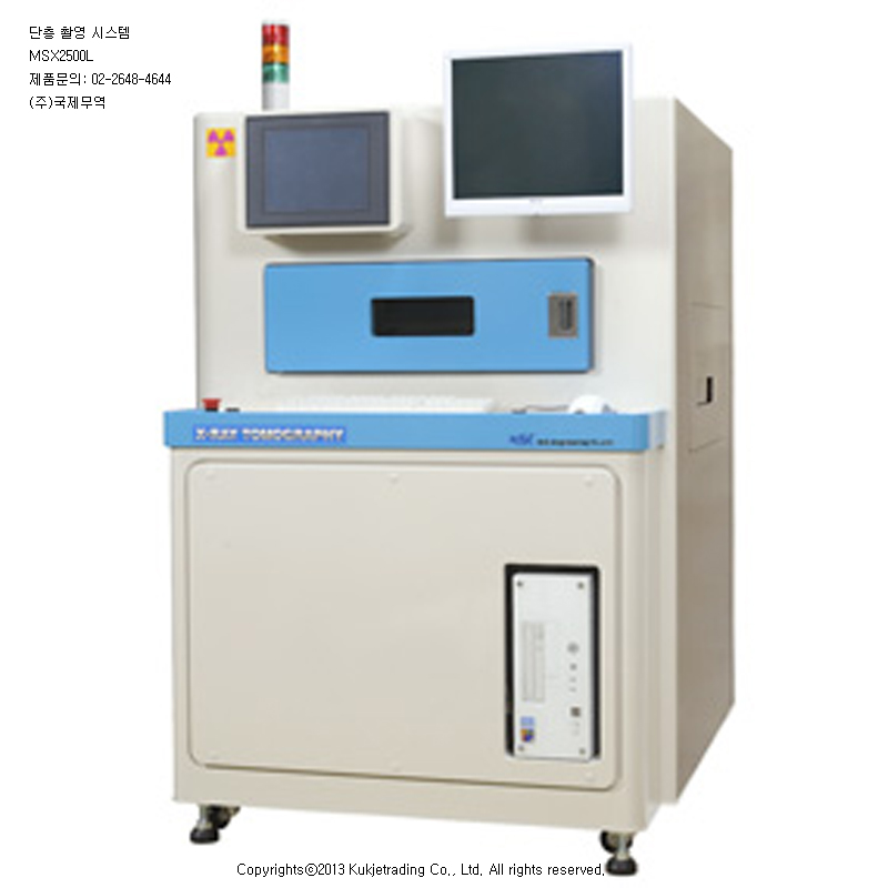
MSX2500L Tomography Inspection System
Feature:
Newest Technology; Tomography by lamina-graph
system.
For Large Size Board; Tomography by lamina-graph
system.
High Speed;The time of a tomogram is 1 seconds
slightly.
High Resolution: The accuracy of a image is 25
microns or more.
Low Price; The price is reasonable
Easy Maintenance:
The X ray generator is closed type with long life.
Semi-In-line possible:
by loader un-loader.
Both-Sided:
Inspection is possible to piled BGA.
Multi Layer Check:
The check of the multilayered board is possible.
Abstract:
MSX2500
is
tomography inspection system by lamina-graphy, It is the best inspection system
to the multi-sided mounting board with high density. The system is able to
operate like in-line test machine by linking conveyor system, such as board
loader and un-loader.
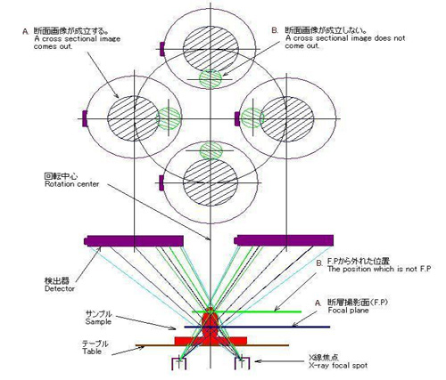
The
lamina-graphy is the theory which synchronized two items in from three items
such as a X ray generator, a detector, and a test material.MSX2500 is
synchronized of the X ray generator and the detector, and there are rotated
within around 1 second. and also they themselves rotate. If it does so, a image
focusing will be fixed, and the fixed position of the focus can be change by the
relational position change of the two items.
The accuracy of the image is 25 micron or less when the image area is 10 x 8mm.
and the position of FP can be chosen in a height of within 13mm.
MSX2500L
MSX2000M Lamina-Graphy System
Information of the X Ray:
The
X ray is an electromagnetic wave with short wave-length, it can pass through
between an atom and atoms. The wavelength of visible is about several microns,
but the x ray is about Å. and a penetration is in inverse proportion to the
density. Generally, It is managed by the rule of the Government. It is because,
the x ray gives a physiologically effect to a live-body.
MSX2500 is managed of the radiation less than 1 micro Sv/ Hours / cm. It is
lower than the color television, the CRT of the color television is always
emitting the several micro Sv/H of radiation. and also there is same levels of
radiation in the atmosphere always.
The radiation of MSX2500 is controlled by the very safety level. It is very
safety zone. Furthermore, MSX2500 is used of the X ray in the sealed box. and it
is with sufficient interlock function.
Test operation Images by MSX2500L
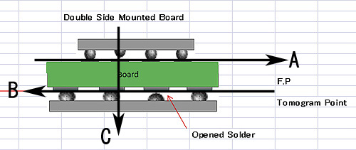
This
is an test sample, in case of two kinds of the BGA is mounted of surface and
reverse-side at the board.
The
tested image of the position B level is displayed on the screen when the FP of
the tomogram is setup as B level.
And
also test image of the position A will be displayed on the screen when the FP is
setuped as A level.
The
tested image of the C will be displayed when tested by the fluoroscopy system.
In this case, inspection will be impossible by the image A and B overlaps.
The
test position of the FP can be adjust in the height of a maximum of 13mm from
the surface of the test table.
.
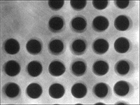
The test image at A.
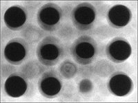
The Test image at B
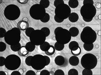
TheTest image by C
Dimensions:
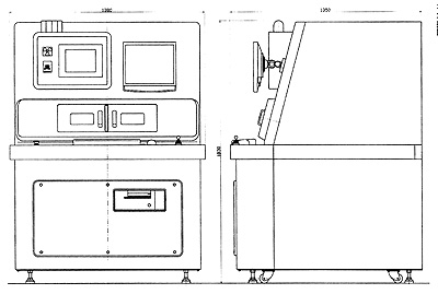
MSX2500L: 1300W x 1600H x 1550Dmm
Semi-In-Line System:
MSX2500 can be check with high speed, it will be inspection around 1 second by
10 x 8 mm area. And also the system can be test by the programmable XY automatic
controller. Therefore, it can operate as automatic X ray tomography inspection
system by with the optional loader with the un-loader system.
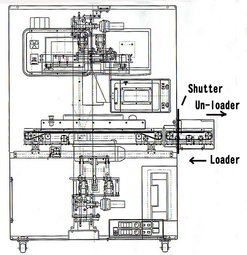
MSX2500:semi-
In-Line type Tomography System
Specifications: Tomography Inspection System MSX2500L
|
Items
|
Specifications |
|
Board Size
|
470X x 540Y mm
max |
|
XY Table
Stroke |
250Xx350Ymm |
|
XY Table
Control |
Motor
Controlled by touch panel |
|
XY Table
Size |
470W x 540D mm |
|
Position
marker |
Red marker by
laser beam |
|
X Ray Source
Voltage |
100 Kv
20W max. |
|
X Ray Tube
Current |
200 micro A
max. |
|
Focus Size
|
5 micron m |
|
X Ray Tube
|
Enclosed
Reflection Type |
|
Cooling |
Air cooling
Closed |
|
Image Angle
|
0 |
|
X Ray
Radiation |
Less than
1µSv/H |
|
X Ray
Protection |
X Ray protected
special Cabinet |
|
Image
Processing software |
GtAVision |
|
Image
Magnification by Tomography |
20X
(25/30/35/40 by Digital Image) |
|
Detector
(Flat Face) |
FOV 1.5 inch |
|
CPU |
3.1GHz
Intel Core |
|
Memory |
4GHB |
|
Storage Unit
|
HDD 1st/80GB
2nd/1TB
DVD |
|
OS |
Windows XP
SP3 |
|
LCD |
17 inch LCD |
|
Image
Controller |
Quality Control
for Averaging, Integrating. |
|
Enhancement
Processing |
|
Contrast &
Brightness Control |
|
Tomogram |
Scanning |
Horizontal
Section |
|
Range of
Height |
0 to 13mm
from
the table |
|
Magnification |
20X (Digital
zoom 25/30/35/40) |
|
Power |
AC230V 800VA
|
|
Body Size
|
1300W x 1720H x
1550D mm
|
|
Weight |
900Kg approx.
|
|